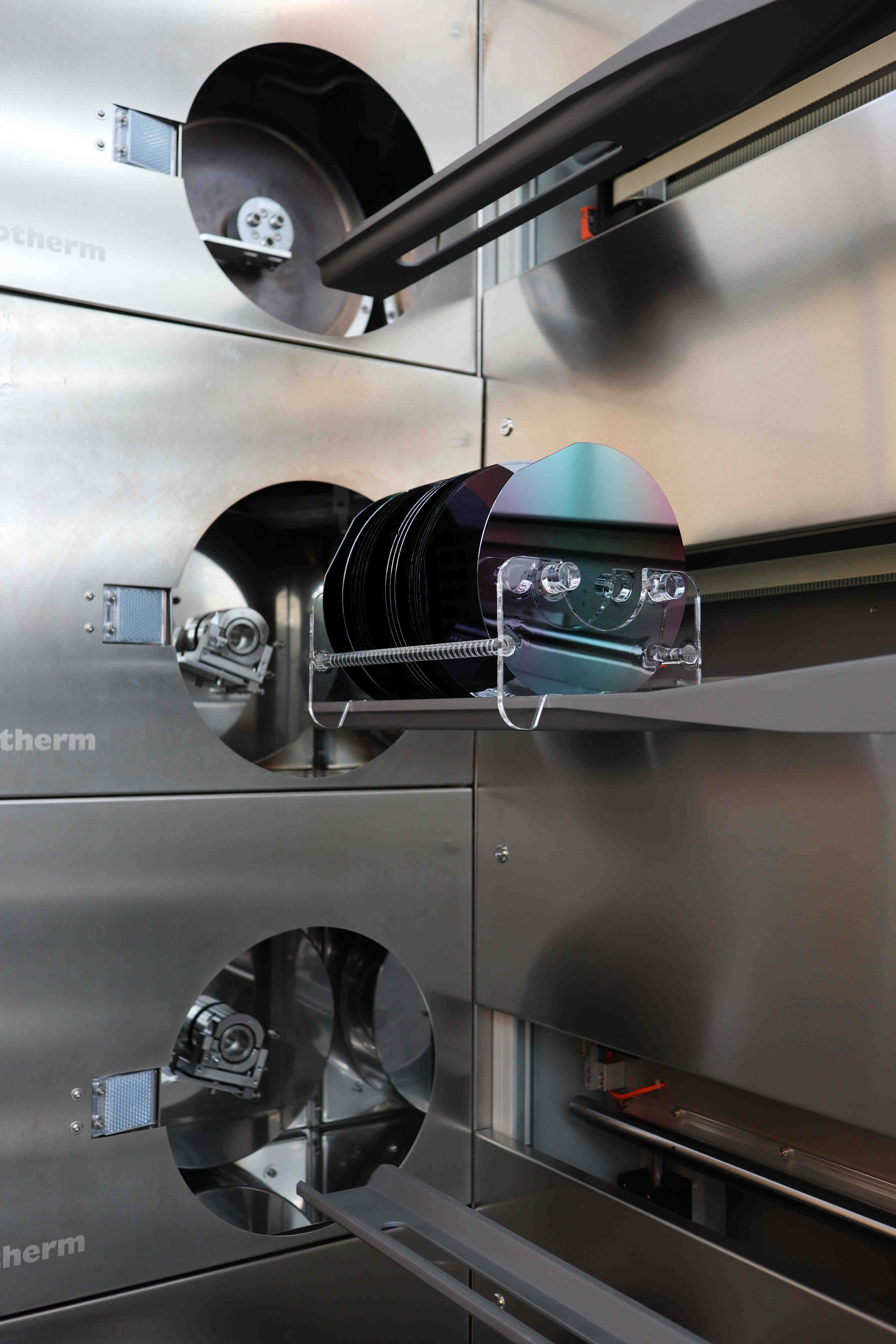
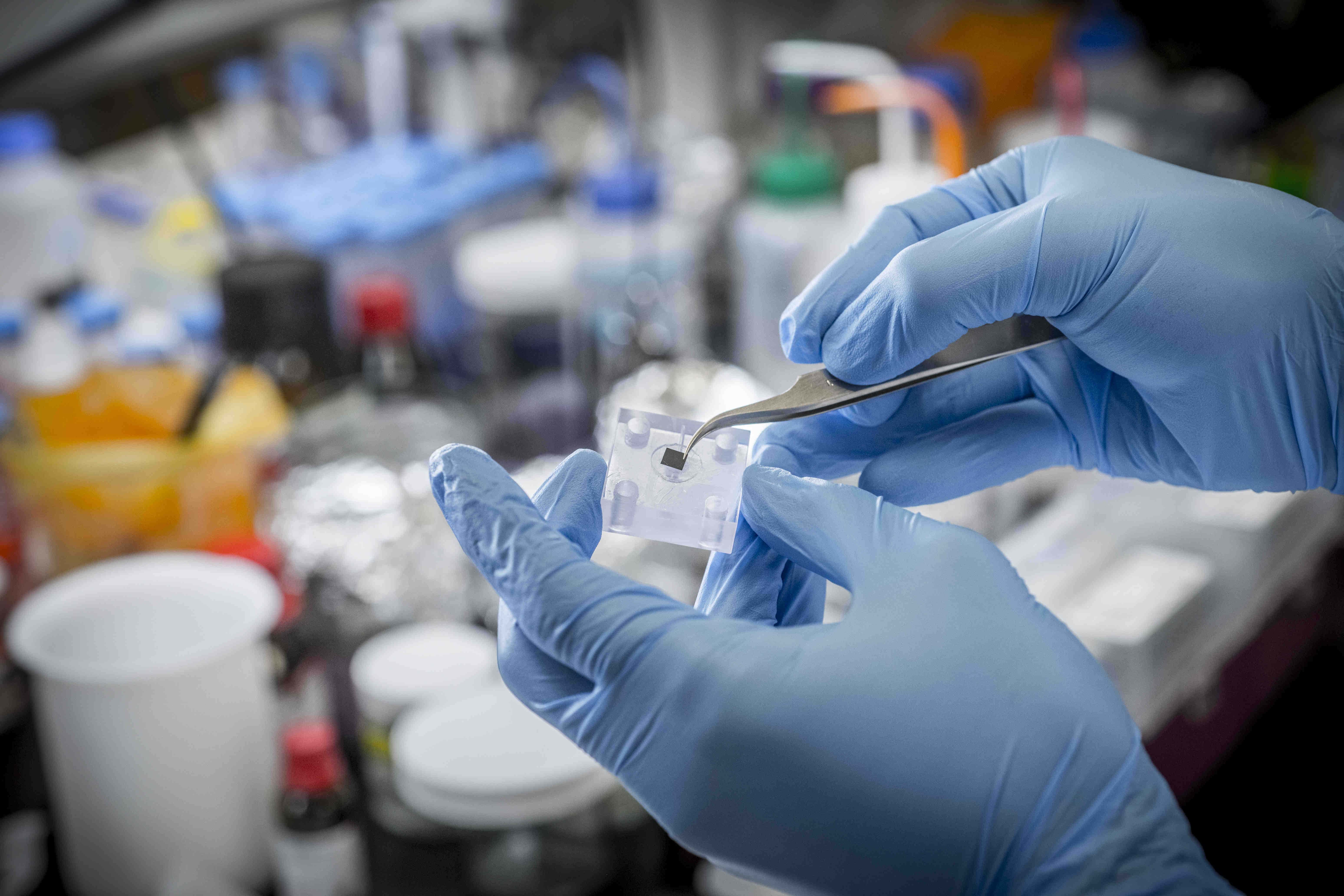
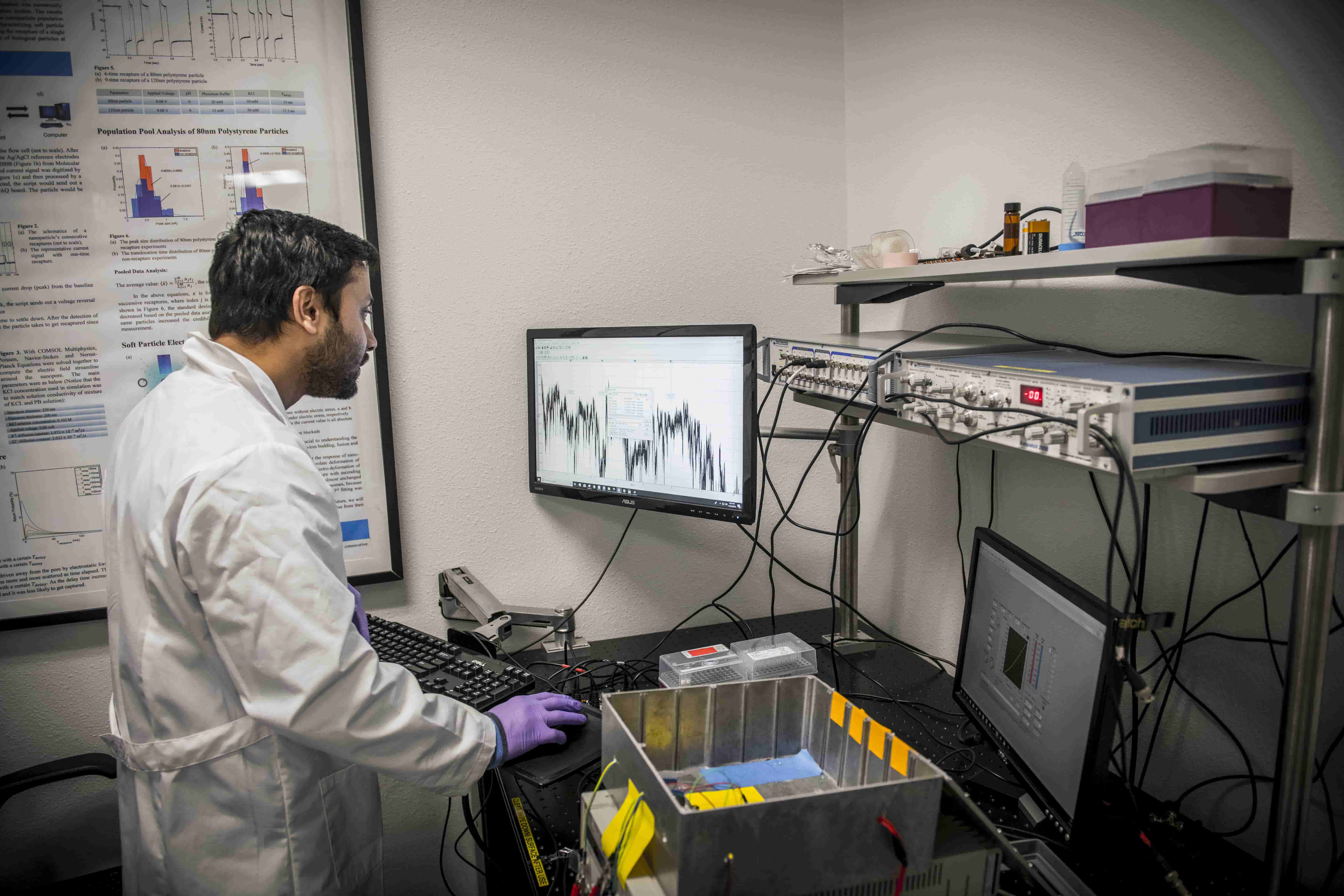
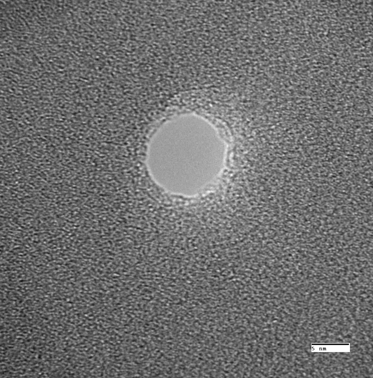
The translocation of analytes through nanometer-sized pores in solid-state or biological membranes has attracted significant attention over the last decade, in both academia and industry. The key idea is to monitor the ionic current across a nanopore, and to associate any transient modulation of it (resistive/conductive pulse) to the translocation of a molecular species through the pore. The resulting “signature” in the current may then be used for probing the biomolecule itself. The aim of our nanopore research is to define a new nanoanalytical technology which will enable efficient detection of conformational changes with microsecond resolution in order to answer a fundamental question about structure and conformations of proteins, DNA molecules, or protein-protein complexes. It is achieved by electrophoretically translocating bioanalytes through a solitary nanopore drilled in thin silicon nitride membrane and analyzing their corresponding current signatures. This has enabled us to study binding-unbinding and folding-unfolding kinetics of single protein molecules. In addition, we have developed DNA sensing platform based on graphene nanopores drilled in single or multi-layer graphene structures which provide exquisite control of the electric field drop within the pore. Nanopore edges are modified with Graphite Polyhydral Crystals (GPC) and the modified GPC nanopores can be used to sense small DNA molecules (down to 25 nucleotide long) which is a significant improvement in the sensing ability of solid-state nanopores. Lastly, we are developing smart solid-state nanopore and nanopore array with superior chemical and mechanical robustness and pore size variability as ultra-fast high throughput nanopore sensors for biophysical characterization of protein-protein interactions and Ag nanoclusters.
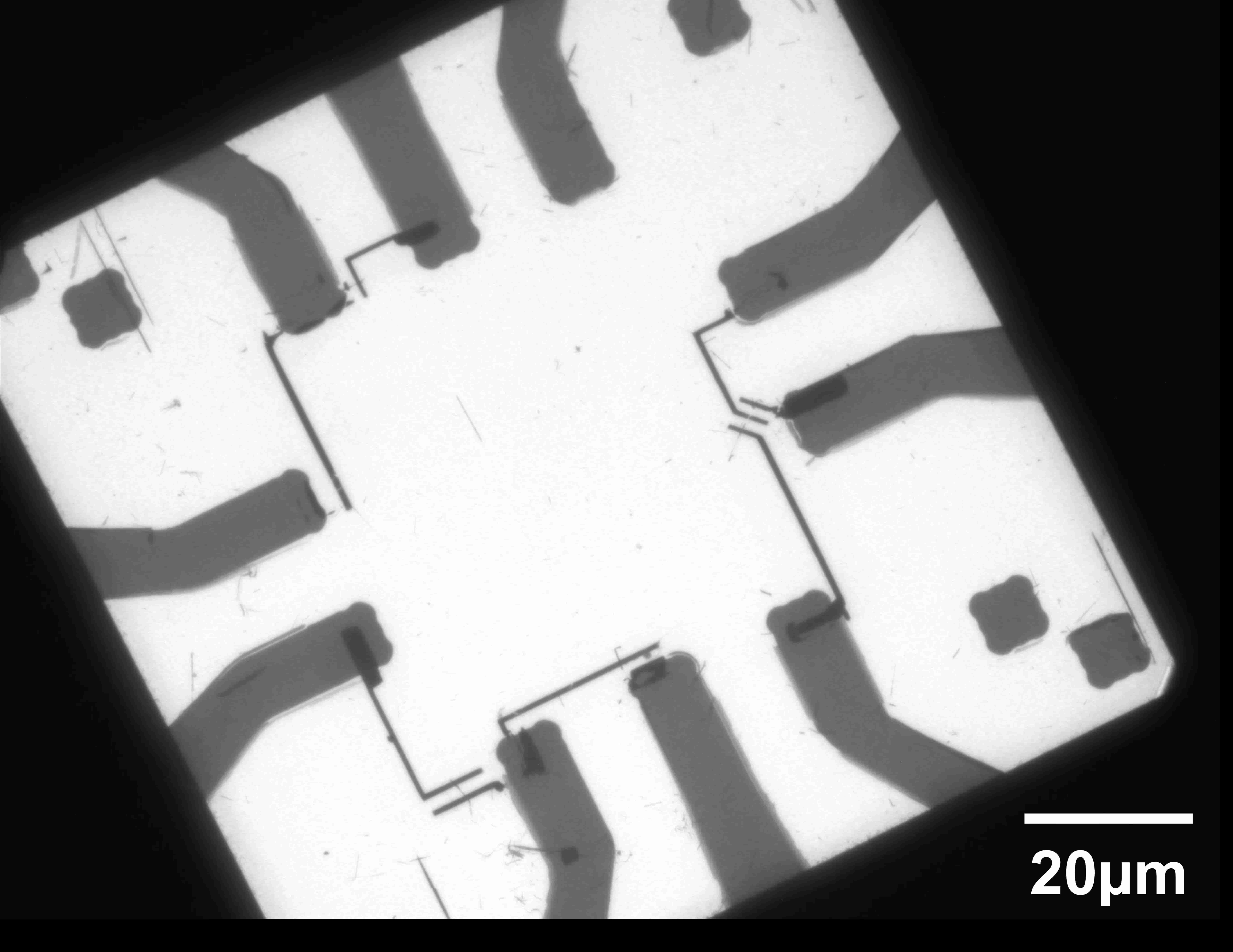
Protein profiling has gained immense attention due to its power to identify disease and therapeutic target biomarkers. In collaboration with Prof. George Alexandrakis at the University of Texas at Arlington, we have developed a stacked nanosensor profiling technology for identifying hitherto unattainable types of biomarkers, complementary to existing assays, by probing single protein and complex dynamics, tether-free and label-free. This new technology consists of two bimodal plasmonic nanopore layers: a first layer for studying temperature-dependent unfolding and a second layer for studying optically and electrically induced unfolding and its effect on protein-receptor (P-R) complex dynamics. The measurement setup is immersed in a constant temperature bath to enable study of protein and complex relaxation and denaturation dynamics above and below room temperature. Both sensor layers are based on our novel self-induced back action (SIBA) actuated nanopore electrophoresis (SANE) concept, which allows study of protein interactions at concentrations 1000-fold below the bulk equilibrium dissociation constant (kd), making this technique ultra-sensitive. We use human serum transferrin protein (hSTf) and its receptor in proof-of-concept studies to show that our nanosensor can differentiate between hSTF species that differ by the amount of iron they carry, which is important as an iron deficiency biomarker. Bimodal optical-electrical signatures have been established for each of the hSTf species for selective admission of bound complexes over unbound proteins in a mixed solution to enable us to deploy the sensor in a complex matrix environment. By modulating laser power, we are investigating the effect of thermal unfolding on different hSTf species and their P-R complexes. Subsequent recapture in a plasmonic trap in the second sensor layer will be optimized for analyte concentrations, applied voltages and nanopore noise. We will use symmetric (VCapture = VRecapture), followed by asymmetric (VCapture ≠ VRecapture) voltage conditions to investigate kinetic parameters associated with P-R complex formation, binding strength and protein relaxation times to differentiate hSTf species kinetics (potential biomarkers) and to study whether voltage-induced protein unfolding is reversible or not – a fact not known yet.
As cancer therapy evolves towards personalized treatments, such as immunotherapy, consideration of a patient’s pre-treatment biomarkers becomes essential. There is growing need for molecular analysis technologies that can verify if a biomarker of interest is indeed targeted by the proposed immunotherapeutic. An emerging trend in novel and very promising immunotherapy interventions involves the targeting of Major Histocompatibility Complex (MHC) class I molecules, also termed Human Leukocyte Antigens (HLA), with recombinant T-cell receptors (TCR) and recombinant TCR-like antibodies in different druggable formats (i.e. bispecific T-cell engager) that mediate specific cancer cell killing. The high-specificity tools needed to assess empirically whether a candidate TCR-like antibody will target peptide-presenting MHC ligands (pMHCs) in a patient’s tumor are not readily available. New technology is needed that can identify antibody binding to limited copy number pMHCs (10-100 per cancer cell) in miniscule (nanoliter) analyte volumes to enable multiple antibody testing with minimum available biopsy. LC/MS/MS can identify pMHCs, but it is expensive and cumbersome to use for target quantification. Flow cytometry, ELISA and functional assays like ADCC and CDC can help identify antibodies that bind to pMHCs but are challenged by lower copy numbers and tumor heterogeneity. Most current protein-ligand interaction quantification technologies, whether using label-free plasmon resonance (e.g. Biacore) or contrast agents (e.g. protein microarrays), suffer from non-specific binding to surfaces of immobilized targets, which impedes their clinical translation. Immunohistochemistry is a good validation tool but requires significant amounts of tumor biopsy (>100 mg) that are often not available from the patient. Single cell RNA-Seq and proteomic approaches were also recently proposed to identify peptide-specific TCRs, but these require further development to meet the demands of personalized clinical applications. Targeting of a limited number of pMHC ligands (10-100) presented per cancer cell by TCR-like antibodies can induce a potent antitumor response. However, quantifying these sparse and heterogeneously expressed ligands from minimal biopsy tissue amounts is not feasible with current technologies. In collaboration with Prof. George Alexandrakis at the University of Texas at Arlington, we are trying to demonstrate feasibility for a label-free sensor to detect these low copy number targets using only a few thousand cancer cells per assay. Superb sensor sensitivity is attained by optical trapping that prevents protein complexes from translocating quickly through a nanopore located at its center and enables simultaneous quantification of size (optical signal) and effective charge (electrical signal) as independent measurements to detect complex formation. The sensor will be integrated with microfluidic isotachophoresis (ITP) technology to facilitate simultaneous concentration increase and enrichment of targeted complexes prior to reaching the sensor. In our studies, we are using TCR-like antibody-pMHC complexes enriched from cancer cells and human tumor xenograft (PDX) tissues to estimate pMHC copy number and compare sensor sensitivity with conventional assays. Successful demonstration of this technology will be a first step towards an instrument that would enrich aspiration biopsies on-chip for pMHCs, screen them with a TCR-like antibody library to quantify tumor heterogeneity in targeted pMHCs and guide therapeutic antibody selection.
The extraordinary chemical and physical properties of biomolecule-templated noble metal nanoclusters along with their potential as the next-generation contrast agents, molecular sensors and logic-gate devices warrant them to be prime candidates for the development of novel bio-nanomaterials. Noble metal nanoclusters, for example, silver (Ag) nanoclusters consisting of ~2-20 Ag atoms conjugated with 10-30 nucleotide long DNA strands are intriguing fluorescence nanomaterials with high extinction coefficient, large fluorescence quantum yields, high photostability, low cytotoxicity, and limited blinking. More importantly, clusters’ stoichiometry, geometry, charge, and ligand-cluster binding orientation profusely influence their optical spectral properties. Therefore, DNA-encapsulated Ag nanoclusters (DNA-Ag-NCs) are a unique type of fluorescent reporter whose fluorescent emission spectrum can reflect subtle changes in its surrounding nucleobases. If carefully designed, these DNA-Ag-NCs can function as a multicolor probe for single-nucleotide polymorphism and N6-methyladenine (6mA) detection. While DNA-Ag-NCs are truly amazing molecular probes and what we have discovered is just the tip of an iceberg, there is no predictive way to design Ag-NCs with desired photophysical properties for various applications. In collaboration with Prof. Tim Yeh at the University of Texas at Austin, we design and create integrated characterization platforms to investigate DNA-templated Ag-NCs. Combined with advanced analysis tools and deep learning models, we aim to elucidate the design rules for producing noble metal NCs with defined architectures and properties.